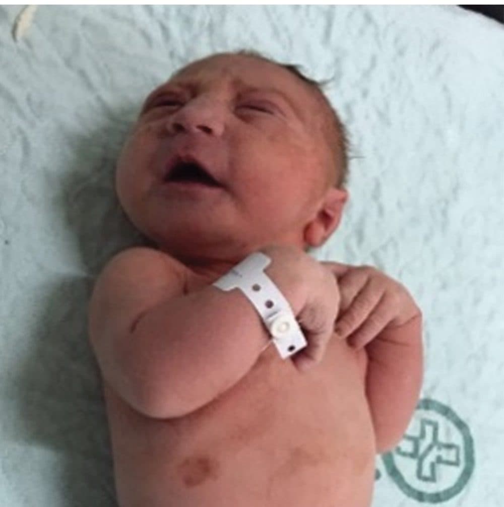
Case of a 34-year-old woman with confirmed Zika virus infection, initially seen for a rash at 8 weeks of gestation. Photograph of the neonate after birth. (Courtesy of Radiology, Radiological Society of North America)
Boston researchers have released the most complete picture so far of how the Zika virus can damage the brain of a fetus.
Their study, published in the journal Radiology, shows detailed graphic images of how the virus can affect the developing brains of babies.
The report suggests that microcephaly — a condition that results in babies born with brain damage and unusually small heads — could be just one of the things that Zika can do to the developing brain.
Guest
Dr. Deborah Levine, director of Obstetric and Gynecologic ultrasound at Beth Israel, and a professor of radiology at Harvard Medical School. Co-author of the Radiology study.
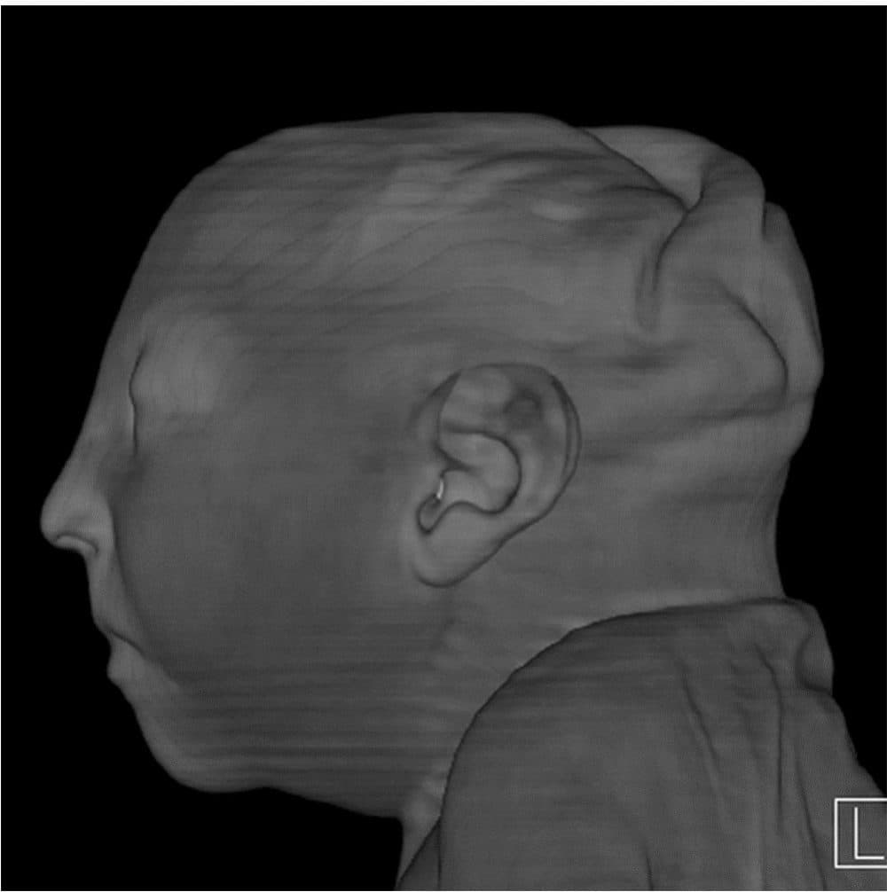
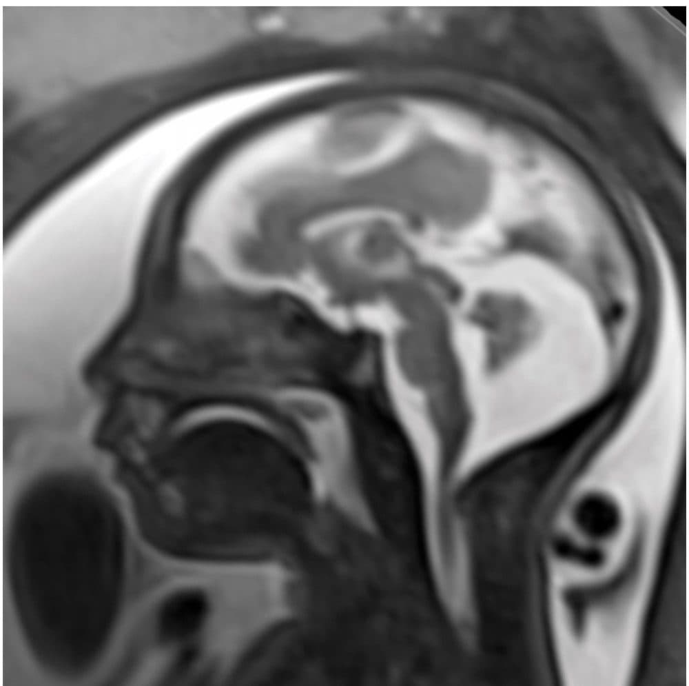
Image obtained at 37 weeks in the case of an 18-year-old woman, first seen for rash at 10 weeks of pregnancy, with confirmed Zika virus infection. (Courtesy of Radiology, Radiological Society of North America)Surface reconstruction postnatal CT image obtained 1 week after delivery at 38 weeks of gestational age in the case of a 24-year-old woman pregnant with twins, with characteristic rash at 9 weeks of pregnancy and confirmed Zika virus infection. (Courtesy of Radiology, Radiological Society of North America)Image obtained at 29 weeks in the case of a 34-year-old woman with confirmed Zika virus infection, initially seen for a rash at 8 weeks of gestation. Sagittal fetal MR images show atrophic frontal lobes, wide sylvian fissures, enlarged posterior fossa, abnormal gyral pattern, prominent cerebrospinal fluid spaces, and inferior vermian hypoplasia. The hypoplastic corpus callosum can be seen, as well as the inferior vermian hypoplasia, enlarged cisterna magna, and heterogeneous signal intensity in the confluence of sinuses. There is a subjectively thin spinal cord. (Courtesy of Radiology, Radiological Society of North America)
Deborah BeckerHost/Reporter Deborah Becker is a senior correspondent and host at WBUR. Her reporting focuses on mental health, criminal justice and education.