Advertisement
Diaper Power: Expanding Gel Could Help Scientists See Brain Workings
If this were a glam-genius movie along the lines of "The Imitation Game," we'd see an exhausted, stymied scientist changing the over-wet diaper of his cranky baby, then suddenly straightening up and gasping in the throes of a revelation: "What if — what if — we don't try to improve the microscope? What if we just make the thing we're trying to see bigger? We could expand it just like the gel in this huge wet diaper!"
Sadly, it didn't happen that way. So the moral of this story is not that scientists should change more diapers. But a report just out in the journal Science does point the way to a promising new scientific tool that could prove helpful in the monumental efforts under way to map the brain. And yes, it involves diapers — or rather, the polymer gel that makes disposable diapers expand so rapidly when wet.
Turns out, with some chemical tweaking, that gel can be used to expand brain tissue without distorting its structure, so it may allow scientists to map the nano-scale 3-D connections between neurons — even potentially to get a full picture of how information flows in small animal brains or parts of the human organ.
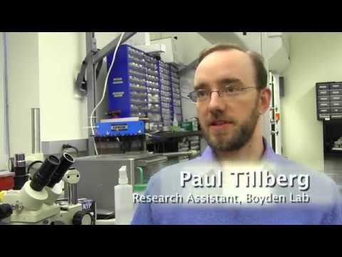
I spoke with neuro-engineer Ed Boyden of MIT's Media Lab and McGovern Institute for Brain Research, senior author on the new study in Science, co-authored with MIT grad students Fei Chen and Paul Tillberg. Our conversation, lightly edited:
How would you sum up what you report in this "Science" paper?
Over the last several hundred years, microscopists have been imaging life. The way they do it is they use a glass lens to magnify the light coming out of the biological sample. This has been very, very powerful, and untold numbers of insights have emerged from it, but there’s a problem: How can you image a large, 3-D object with nano-scale precision? Light cannot go down to very, very fine precision because light is sort of finite in size, you could say. It has a wavelength that’s really large compared to single molecules.
What we’ve found is that, in contrast to lens-based magnification, you can physically magnify an object and make it bigger. So that was the first key finding: We can physically magnify an object.
The second key finding is that we have engineered a chemical system that lets you do this very, very precisely and with good resolution.
And a third take-home message from the paper is that we have now shown that the chemical process we developed is very isotropic — that is, it’s very smooth and even, and doesn't introduce distortion, all the way down to the nanoscale.
Why does being able to analyze brain tissue at this nanoscale resolution matter?
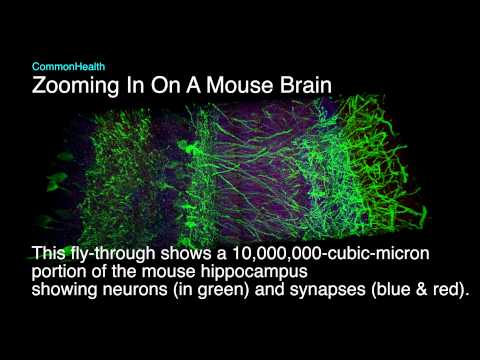
If you want to understand the brain, well, brain circuits are quite large. The individual cells in the brain could be millimeters or centimeters in size in terms of their length. But the actual things that organize the brain — the connections called synapses — are nanoscale. So if you want to understand how a brain circuit funnels information or processes information, you need to be able to map a large, 3-D object with nanoscale precision, and that's something our technology is enabling.
What could be done with it?
In neuroscience, we’re excited by the possibility that you could try to map an entire small brain, in organisms like flies or worms. We think it's possible you could expand the entire nervous system or the entire brain and then see the whole thing.
That would be very exciting because you could try to follow the pathways that lead in from the sensory organs — like the eyes — all the way to the motor outputs — to the muscles, and look at all the stuff in between: What makes decisions? What makes memories? And then map that.
One could imagine that at some time in the future you could try to load up these molecular maps of a neural circuit into a computer and then try to simulate a brain in a computer.
What about human brains?
Human brains are really large. If you were to map an entire human brain with this sort of single-protein resolution — this is a back-of-the-envelope estimation — if you were to store that on hard drives and you stacked all those hard drives, one on top of the other, with reasonable resolution and enough information about each point in the brain, that stack of hard drives would reach into outer space.
So I think what will happen is that we can map parts of the human brain. A couple of our collaborators are starting to explore applying expansion microscopy to human brain tissues, and that’s a work in progress now. But in the short term, in terms of mapping an entire brain, it will probably have to be small model organisms that are common in basic and applied neuroscience.
What will your own lab do with this technique?
I think we’re going to expand the entire brain of small animals, like flies, and see if we can actually see how all the neurons look: Can we actually start to make a parts list for the brain? Can we start to look at the connections and figure out which ones are strong or weak, which ones are fast or slow, and do it across an entire neural circuit? I think the data we get could be very useful for trying to understand how information flows in the brain.
Was there a sudden moment of thinking "out of the box," when someone said, "Instead of improving microscopes, what if we make the sample bigger?"
We’ve been talking for quite some time in our group about making tissues bigger so you can just image them with nanoscale precision, but it was almost like a joke or something you'd say out of desperation when you were frustrated with a regular microscope.
But there was a point in time a couple years ago, probably in mid-2012, when we were really trying to do nanoscale imaging using strategies including some related to the technologies that won the Nobel prize in chemistry last year. And they were very difficult to use, especially for large, 3-D samples. So then we started thinking more seriously: Well, what if we actually did make it bigger? That’s when we stumbled across a series of papers actually dating back many decades, where people were studying this material very much like what's in baby diapers and showing that it can indeed undergo these controlled, dramatic changes in size.
Without changing the structure in any way that matters?
When we tried out the polyeletcrolyte gel to see if we could expand brain tissues, it didn’t work completely right away. We were able to get expansion but sometimes it would cause distortion or cracking. So to actually get the chemistry to work well took a couple years of trying out new chemistries and carefully validating them by comparing them to earlier technologies and so on. But now it works pretty well at a resolution of about 60 nanometers, which is quite small.
It can expand the sample by a factor of four or five — is that really enough?
An off-the-shelf conventional microscope, due to the limitations of the wavelength of light, can image things down to about 300 nanometers or so. That's hundreds of times bigger than an individual biomolecule. We'd like to close that gap between what microscopes can do and what biology and medicine really need. So right now we have a factor of 4 or 5 in terms of improvement and so that means we’re down to about 60 nanometers or so. That's now a scale at which you can actually start to look at these structures inside of cells with great precision. For example, all of the connections between neurons — the synapses — are organized with precision around this range. The gap between two neurons is probably a little bit bigger than that in terms of the size distribution.
I do wish this could be a story about how you gained inspiration from the diapers you changed as a father...
That would be a great story if it were the case, wouldn’t it? We actually stumbled across this in part because the material in the diaper, this polymer, is also just really well studied by physicists, and it's very well understood. We wanted a way to blow up tissues to make them much bigger that would still preserve all the nanoscale architecture. This is a very well studied polymer but it also — very coincidentally — happens to be the same stuff as in diapers...