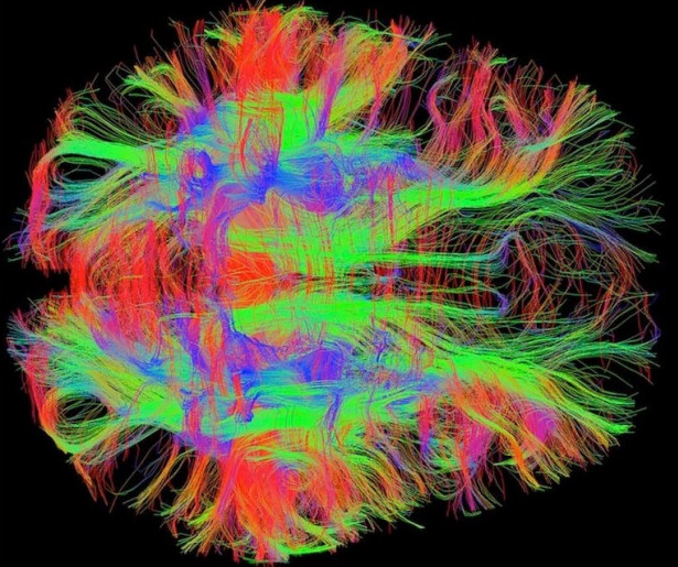
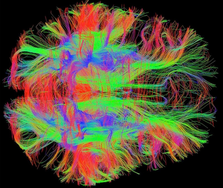
A map of nerve fibers in the human brain (. (Courtesy of Zeynep Saygin/Massachusetts Institute of Technology.)
Happy almost 2015. Instead of doing our usual "Top 10 CommonHealth stories of the year" post, we've decided instead to look back at our tip-top, far-and-away #1 organ of the year for 2014.
Hint: It's well above the waist. The brain is, to quote Pink Floyd: "All that you touch/All that you see/All that you taste/All you feel./All that you love/All that you hate/All you distrust/All you save."
Etcetera. The brain is also the focus of some of the most fascinating research in modern-day science.
Our 2014 series, "Brain Matters: Reporting from the Front Lines of Neuroscience," tried to capture a partial snapshot of this pivotal moment in brain science, a time of new tools and insights so promising that scientists themselves are saying this is the most exciting time ever to work on the brain.
The series included the set of gorgeous images below, compiled by former intern Suzanne E. Jacobs, and a collection of short video interviews with young neuroscientists, produced by WBUR's Jesse Costa: 11 Young Neuroscientists Share Their Cutting Edge Research.
The individual "Brain Matters" pieces, in reverse chronological order:
Wishing you a wonderful new year. Special thanks to WBUR's Iris Adler, who supervised the "Brain Matters" series. And now, for your visual pleasure, the wondrous view inside your head:
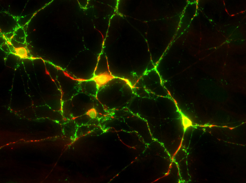
These are cells in a developing rat brain. Neurons are labeled in red, and the connections between them are in green. This image was taken after 21 days of in vitro growth. (Courtesy of Neville Sanjana/McGovern Institute for Brain Research at MIT)
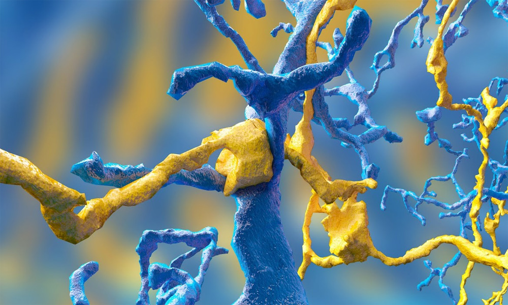
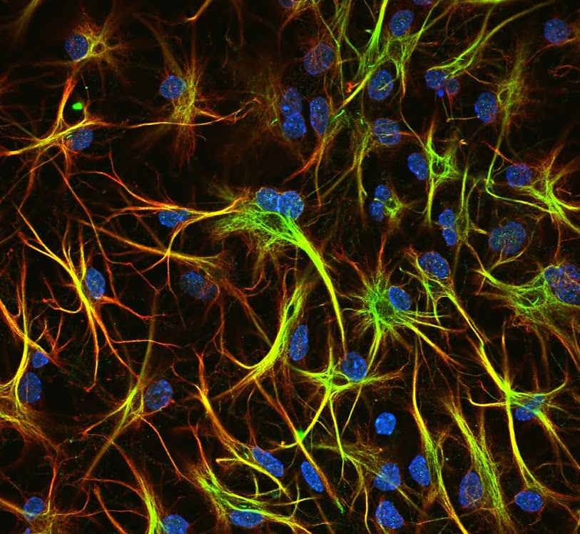
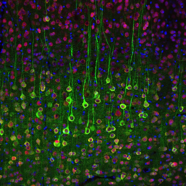
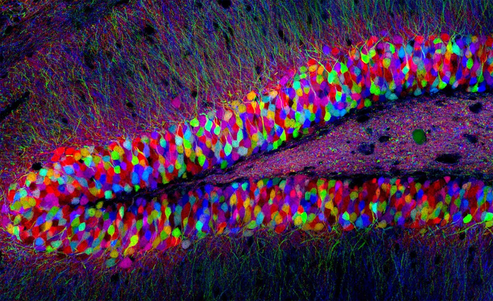
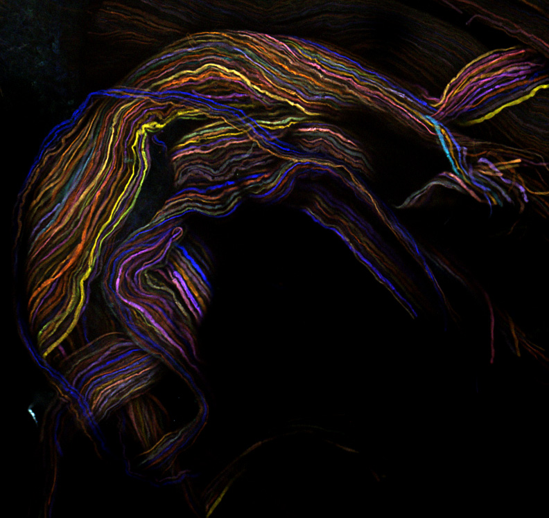
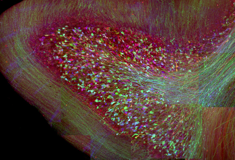
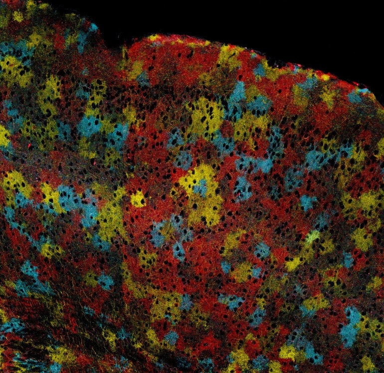
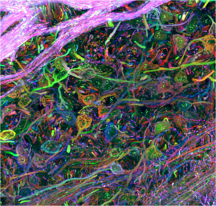
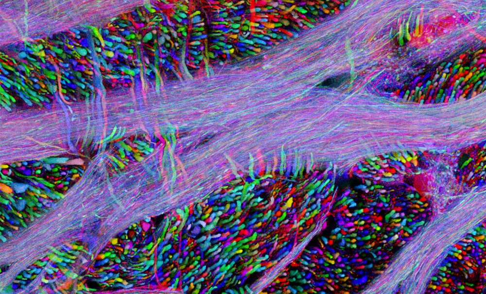
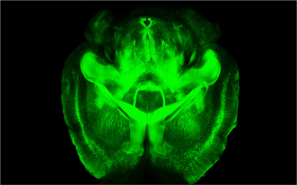
This computer-generated model shows the connection between two neurons in a mouse retina. The image comes from an online game called EyeWire, a citizen-science effort that recruits volunteers from around the world to help build a three-dimensional map of the neural connections in a mouse eye. Sebastian Seung -- a neuroscientist formerly at MIT and now at Princeton University -- launched the game in 2012. In this picture, the blue cell is known as a Ganglion cell, and the yellow cell is known as a Starburst Amacrine cell. The Seung Lab hypothesized in a recent paper that Starburst Amacrine cells are involved in detecting the direction of motion. (Courtesy of Alex Norton/EyeWire)These are mouse neurons that researchers in Guoping Feng’s lab at the Massachusetts Institute of Technology have stained to reveal a protein related to autism and other brain disorders. (Courtesy of Guoping Feng, Michael Wells/McGovern Institute for Brain Research at MIT)These are neurons in a mouse brain. The mouse has been genetically altered so its neurons express a protein that emits light when the neurons are active. (Courtesy of Guoping Feng,Louis Tee/McGovern Institute for Brain Research at MIT)This is a part of a mouse brain called the dentate gyrus. It is located in the brain’s hippocampus, a region involved in memory formation. This mouse was genetically engineered to express fluorescent proteins of varying color combinations in its neurons. This labeling process, called Brainbow, allows scientists to more easily distinguish individual cells and connections in the brain. (Courtesy of Livet, Weissman, Sanes and Lichtman/Harvard University)This is a group of mouse axons: long projections that stem from neurons and carry electrical impulses. These axons are bundled together to form a nerve. This mouse was genetically engineered to express fluorescent proteins of varying color combinations in its neurons. This also used the Brainbow labeling process. (Livet, Weissman, Sanes and Lichtman/Harvard University)This is the cross-section of a mouse cerebellum, a brain region involved in motor control. This mouse was genetically engineered to express fluorescent proteins of varying color combinations in its neurons.This also used the Brainbow labeling process. (Livet, Weissman, Sanes and Lichtman/Harvard University)This image shows cells in a mouse brain. The black spots are neurons, and the colored cells between the neurons are astrocytes, the most abundant cell type in the brain. Among their many functions, astrocytes provide structural support to the brain, fuel neurons, and form the blood-brain barrier. This mouse was genetically engineered to express fluorescent proteins of varying color combinations in its neurons. This also used the Brainbow labeling process. (Livet, Weissman, Sanes and Lichtman/Harvard University)These are cells in the brainstem of a mouse that was genetically engineered to express fluorescent proteins of varying color combinations in its neurons. This also used the Brainbow labeling process. (Livet, Weissman, Sanes and Lichtman/Harvard University)This is part of the brainstem of a mouse that was genetically engineered to express fluorescent proteins of varying color combinations in its neurons. This also used the Brainbow labeling process. (Livet, Weissman, Sanes and Lichtman/Harvard University)This is a mouse brain that scientists made transparent using a new technique called CLARITY. First, they infused the brain with a gel that bound to the brain's proteins and held them in place. Then, the scientists flushed out all the opaque fatty tissue from the brains, leaving them structurally intact but transparent. Using this technique, scientists can study the three-dimensional structure of brains without having to cut them apart. (Courtesy of Deisseroth Lab at Stanford University)brain-matters
